The Importance of Chest X-Rays in Modern Medicine

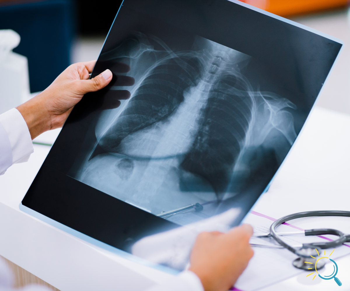
Chest X-rays are one of the most commonly performed diagnostic imaging tests and for good reason. They provide a quick, non-invasive, and cost-effective way to examine the chest’s internal structures, including the lungs, heart, ribs, and diaphragm. Whether you’re dealing with respiratory symptoms, suspected injuries, or routine health assessments, chest X-rays play a vital role in diagnosing and monitoring a wide range of medical conditions.

Why Are Chest X-Rays Performed?

Chest X-rays are typically requested by healthcare providers to investigate symptoms like persistent cough, shortness of breath, chest pain, or fever. They are also commonly used to detect and monitor conditions such as:

  • Lung infections: Pneumonia, bronchitis, or tuberculosis.
  • Heart conditions: Enlargement of the heart or fluid around the heart.
  • Injuries: Fractures of the ribs or other trauma.
  • Lung diseases: Chronic obstructive pulmonary disease (COPD), emphysema, or lung cancer.
  • Other abnormalities: Tumors, fluid buildup, or foreign objects in the chest.

For individuals with chronic conditions, such as asthma or heart disease, chest X-rays are often used to monitor progression and guide treatment plans.

How Chest X-Rays Work

Chest X-rays use a small dose of ionizing radiation to create images of the chest’s internal structures. The process is simple and quick, taking only a few minutes from start to finish. During the procedure, you will stand or sit in front of the X-ray machine while the technician captures the images. These images provide a detailed view of the chest’s internal anatomy, enabling healthcare providers to pinpoint issues that may not be visible through physical examinations alone.

Benefits of Chest X-Rays

One of the most significant benefits of chest X-rays is their ability to provide immediate results. This makes them an invaluable tool in emergency settings where prompt diagnosis is critical. Additionally, chest X-rays are:

  • Non-invasive: There’s no need for incisions or invasive procedures.
  • Low-risk: The radiation dose is minimal, making the procedure safe for most patients.
  • Affordable: Chest X-rays are one of the most cost-effective diagnostic tools available.

Preparing for Your Chest X-Ray

Preparation for a chest X-ray is minimal. You’ll be asked to remove jewelry or clothing with metal components that could interfere with the imaging process. It’s also essential to inform the technician if you are pregnant or suspect you might be, as additional precautions may be necessary.

Where to Get a Chest X-Ray in Deltona, FL

If you’re in need of a chest X-ray, choosing a trusted diagnostic center is essential. At Sunshine Diagnostic Center, we offer state-of-the-art imaging services to ensure accurate and timely results. Our experienced team is committed to providing exceptional care in a comfortable setting.

Explore more about our services for chest X-ray in Deltona, FL and schedule your appointment today. Taking charge of your health starts with reliable diagnostics, and we’re here to help every step of the way.

What is an Imaging Exam For?

An imaging exam allows doctors to visualize the internal structures of the body. These exams help diagnose, monitor, and even prevent health issues. Common reasons for imaging exams include identifying fractures, assessing organ function, and detecting tumors.

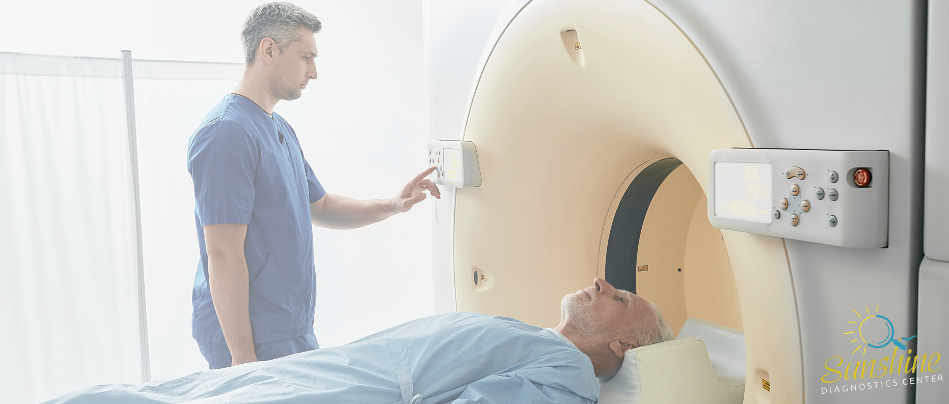
For example, X-ray imaging is often used for broken bones, while CT imaging can provide a deeper look at internal organs and tissues. At Sunshine Diagnostic Center, we offer a range of imaging services to meet your healthcare needs.

What are the Four Types of Medical Imaging?

Medical imaging is vital for diagnosing and monitoring a range of conditions. The four primary types include:

  1. X-ray Imaging: Best for visualizing bones and detecting fractures.
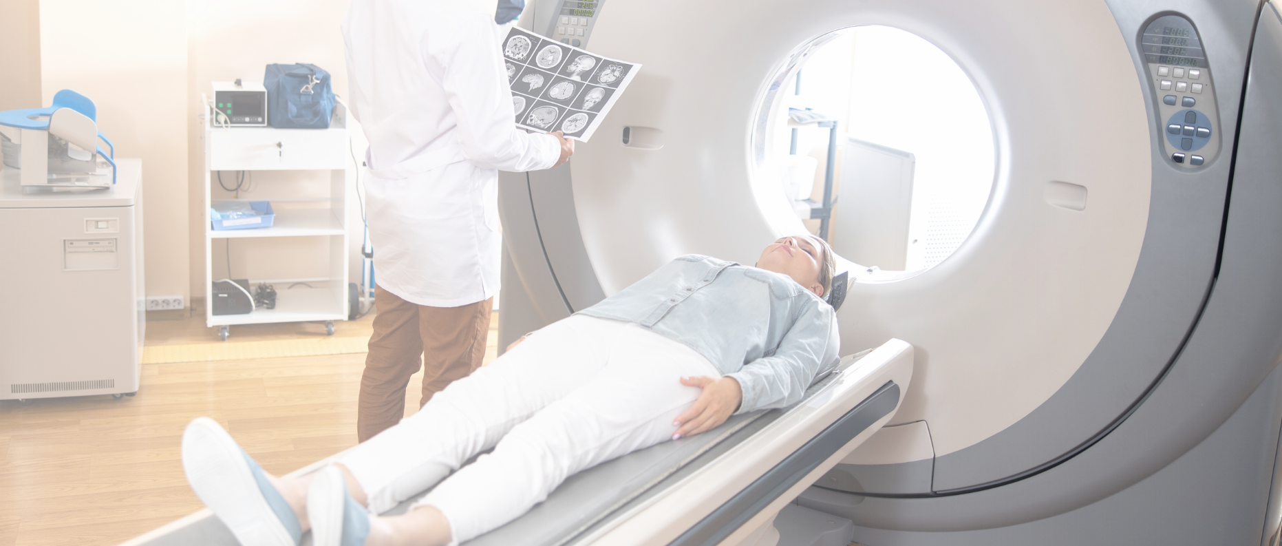
  2. CT Imaging: Produces detailed cross-sectional views of the body, useful for internal organs and complex conditions.
  3. Ultrasound: Uses sound waves to create real-time images, often used for monitoring pregnancies or diagnosing soft tissue conditions.
  4. Nuclear Medicine: Involves radioactive tracers to visualize specific organs and their function.

Each type has unique strengths, depending on the condition being evaluated. Learn more about CT imaging and its versatility.

What is the Difference Between Radiology and Imaging?

Radiology and imaging are interconnected but distinct. Radiology is a medical specialty that uses imaging to diagnose and treat conditions. Imaging, on the other hand, refers to the technologies and techniques—like X-ray imaging, CT scans, and ultrasounds—that create visual representations of the inside of the body.

Radiologists are medical doctors who interpret images to identify abnormalities and guide treatments. Imaging technology is the foundation of radiology, offering critical insights into patient health. For example, X-ray imaging is ideal for detecting fractures, while CT imaging provides cross-sectional views of organs and tissues.

For more on how these techniques come together, explore x-ray imaging at our center.